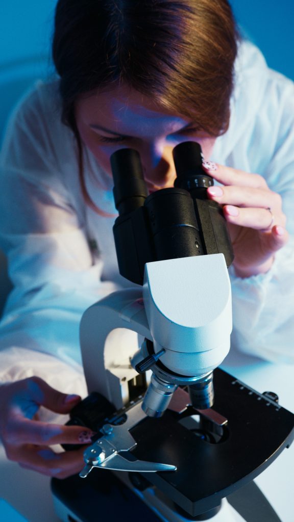
3D CELL CULTURES: IMAGING AND ANALYSIS
Admin / January 24, 2023
Three-dimensional or (3D) cell cultures are useful in cell biology research as well as tissue engineering; as they resemble the architectural microenvironment of natural tissue more closely than two-dimensional cultures. Microscopy techniques function well for thin, optically transparent cultures. On the other hand, they are not accurate when it come to imaging 3D cell cultures. Three-dimensional cultures can be dense and scattering, which prevents light from passing through without significant distortion. Confocal microscopy, multiphoton microscopy, as well as optical coherence tomography are all techniques that can image thicker biological specimens at high resolution. Dynamic changes to cells in 3D microenvironments can be assessed non-destructively over time using these techniques.
2D cell cultures substrates have been known to be the standard technique in the research for cell biology for decades. However, the commonly used culture flasks and dishes are not efficient; as they don’t give replication to the natural microenvironment of cells in tissue. According to research, the extracellular environment has a significant impact on cell biology, including cell differentiation. As a result, there has been a surge in interest in the development of cultivating techniques that are more akin to natural tissue. There are numerous types of three-dimensional objects. Three-dimensional (3D) cultures and scaffold materials have been created.
Three-dimensional cell cultures are becoming increasingly important for research and clinical applications, with one goal being the development of advanced biocompatible engineered tissues. While 3D cell cultures better mimic the natural environment of cells, their use introduces new challenges. Several tools which are used for visualising live cells in 2D cell cultures, rely on light transmission through the sample; these include : field and phase microscopy. These methods are impractical for viewing cells in 3D cultures because the entire sample may be too thick for light to pass through. Imaging techniques that rely on epi-illumination (light collected in the backward direction) are better suited for non-destructive visualisation of 3D cultures and thick tissue samples.
There are various epi-illumination imaging techniques that can detect fluorescence as well as backscattered light. Fluorescence-based techniques are most commonly used to visualise a fluorescent marker that has been specifically targeted to a specific area or molecule of interest. Alternatively, the autofluorescence of cells, as well as tissues, can often provide enough contrast; as to identify both structural and biochemical components. Imaging techniques based on light scattering are commonly used to examine the structural and dimensional properties of a sample. Confocal microscopy (CM) is an imaging technique. It permits for high resolution optical sectioning of relatively thick samples. CM can be used in both fluorescence and reflectance modes. Confocal microscopy has a penetration depth of approximately less than 100 m. Using multi-photon microscopy, deeper penetration depths for fluorescence imaging can be achieved (MPM). Depending on the tissue type, this technique uses nonlinear optical effects to perform high resolution optical sectioning in samples up to a millimetre thick. Optical coherence tomography can achieve scattering-based imaging with penetration depths of several millimetres. OCT creates depth-resolved images by measuring the time-of-flight of scattered photons using interferometric techniques. Choosing the best imaging technique. The optical properties, type, and thickness of the culture, as well as the features of interest in the culture, all influence which imaging technique is appropriate for a specific 3D cell culture.
Some of the imaging techniques can be used concurrently, providing a more complete view of a sample based on different contrast-enhancing mechanisms. These imaging techniques are ideal for observing dynamic changes in 3D cell cultures, as they have very little impact on the viability of living cells.
The samples’ structural and functional properties are visualised using optical coherence tomography, multiphoton microscopy, and confocal microscopy. OCT and CM are used to track the location and functional activity of cells in a chitosin scaffold over several days. High-resolution images of cells seeded on 3D micro-topographic substrates are obtained using OCT and MPM. The Understanding of principles and the limitations of: confocal microscopy, multiphoton microscopy, and optical coherence tomography, is critical during the selection process of choosing the best imaging technique for a particular type of 3D culture, scaffold, or substrate. The optical contrast mechanism (fluorescence or scattering), resolution, and penetration depth are the most important parameters to consider when imaging 3D cultures.
